

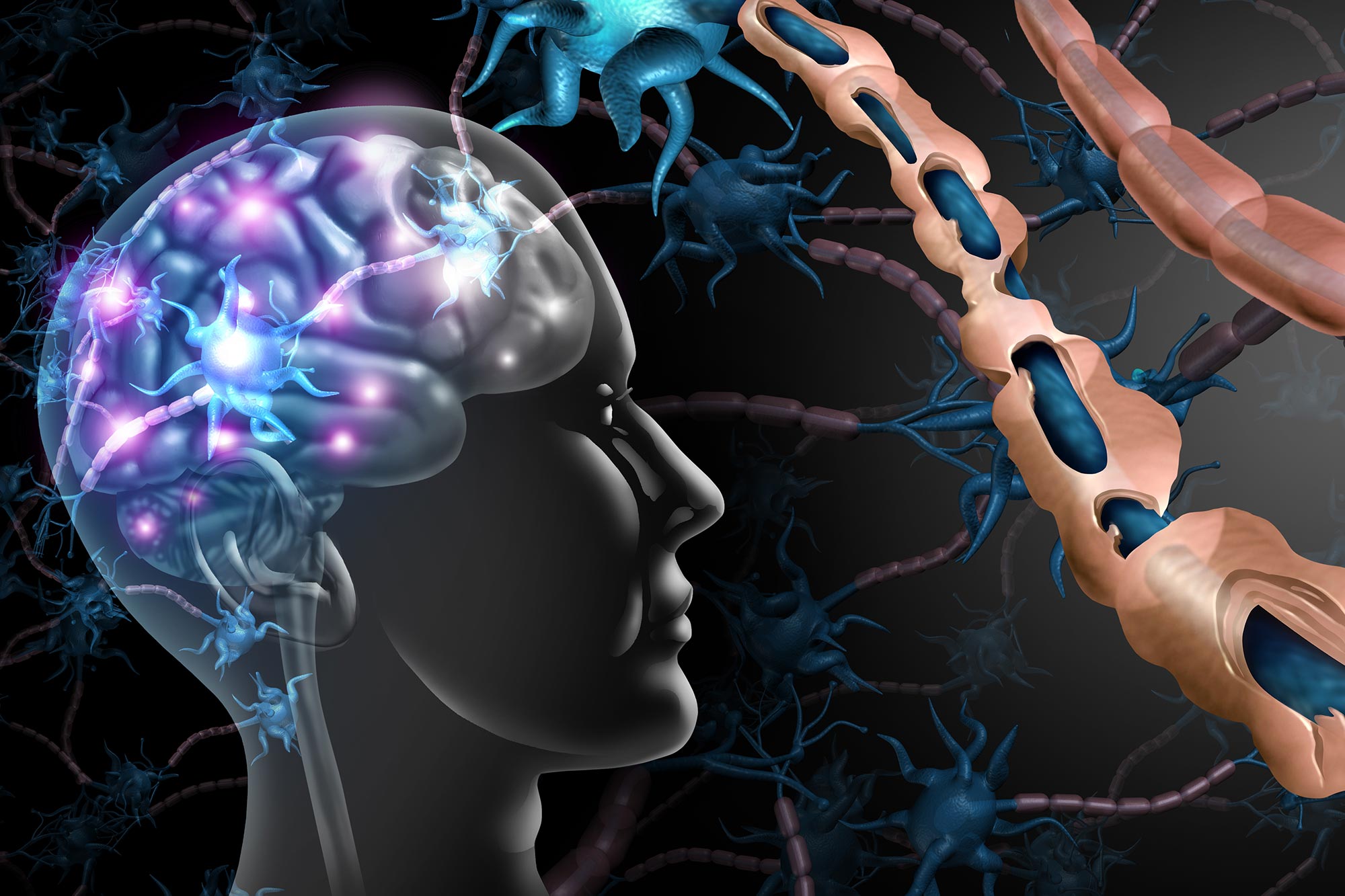
Researchers have developed an artificial intelligence method that can uncover previously hidden signs of multiple sclerosis in routine MRI scans. For decades, multiple sclerosis specialists have been forced to study the disease through an incomplete picture. Standard magnetic resonance imaging (MRI) can reveal damage in the brain’s white matter, but some of the lesions most [...]
Researchers have developed an artificial intelligence method that can uncover previously hidden signs of multiple sclerosis in routine MRI scans. For decades, multiple sclerosis specialists have been forced to study the disease through an incomplete picture. Standard magnetic resonance imaging (MRI) can reveal damage in the brain’s white matter, but some of the lesions most closely tied to disability and cognitive decline have remained largely invisible.
Those hidden injuries occur in the gray matter, particularly the brain’s cortex. Researchers have long known that cortical lesions can offer important clues about how MS progresses and how severely it may affect a patient. Yet routine MRI has not provided a reliable way to detect or monitor them, leaving clinicians with a major blind spot.
Even many of the newer drugs that can slow MS progression are designed primarily around reducing white matter lesions. Now, a University at Buffalo-led research team has used artificial intelligence to expose some of that previously concealed damage in existing MRI scans. Published in Communications Medicine, the study shows how computational methods can compare information across multiple images and recover disease signals that conventional viewing methods fail to capture.
“Detecting previously invisible cortical lesions on conventional legacy MRI scans has major implications for MS research and clinical care,” says Robert Zivadinov, MD, PhD, senior
Source and reference
author on the paper, SUNY Distinguished Professor in the Department of Neurology and director of the Buffalo Neuroimaging Analysis Center (BNAC) in the Jacobs School of Medicine and Biomedical Sciences at UB. “The ability to see for the first time these previously hidden indicators of MS disease progression, including cognitive impairment and disability, is an important advance,” he says. Hidden lesions shaped MS uncertainty Cortical lesions have been linked to MS since nearly the time the disease was first described in the late 19th century. Still, they did not become part of diagnostic criteria until the 21st century, and even then, their usefulness was limited because clinical MRI could not reliably reveal them. “We have all been very frustrated, knowing that these cortical lesions were there but not being able to see them,” says Michael G. Dwyer, PhD, first and corresponding...
Read original source- Published
- Jul 13, 2026
- Updated
- Jul 13, 2026
- Source
- Scitechdaily
- Category
- Health
- Read time
- 4 min
Key facts
Why this matters locally
This health story matters locally because it may affect readers, businesses, commuters, families, or public services in British Columbia.
Local impact
BC Post links this item to British Columbia coverage so readers can follow related city updates, weather, traffic, events, and category news in one place.
Timeline
Source and credit
BC Post may summarize, organize, and add local context for reader clarity. Original reporting remains with the listed publisher.
Related Articles
View all Health
Provation Expands Global Footprint with Launch of Provation® Apex in Australia and New Zealand
Health
Provation Expands Global Footprint with Launch of Provation® Apex in Australia and New Zealand
 Health
Neuroscientists find a teamwork paradox: highly synchronized brains perform worse at complex tasks
Health
Neuroscientists find a teamwork paradox: highly synchronized brains perform worse at complex tasks
 Health
The Surprising Cellular Benefit of the Pigment Behind Red Hair
Health
The Surprising Cellular Benefit of the Pigment Behind Red Hair
 Health
Andlauer-backed group loses bid for Château Montebello
Health
Andlauer-backed group loses bid for Château Montebello
 Health
Coffee May Protect the Liver in More Ways Than Scientists Realized
Health
Coffee May Protect the Liver in More Ways Than Scientists Realized
More Local Coverage
Latest news Business
AI Market Trends: Q2 2026 Review and Forecast
Business
AI Market Trends: Q2 2026 Review and Forecast
 Politics
Powerful pro-life ad featuring boxing legend to air during final World Cup matches
Politics
Powerful pro-life ad featuring boxing legend to air during final World Cup matches