

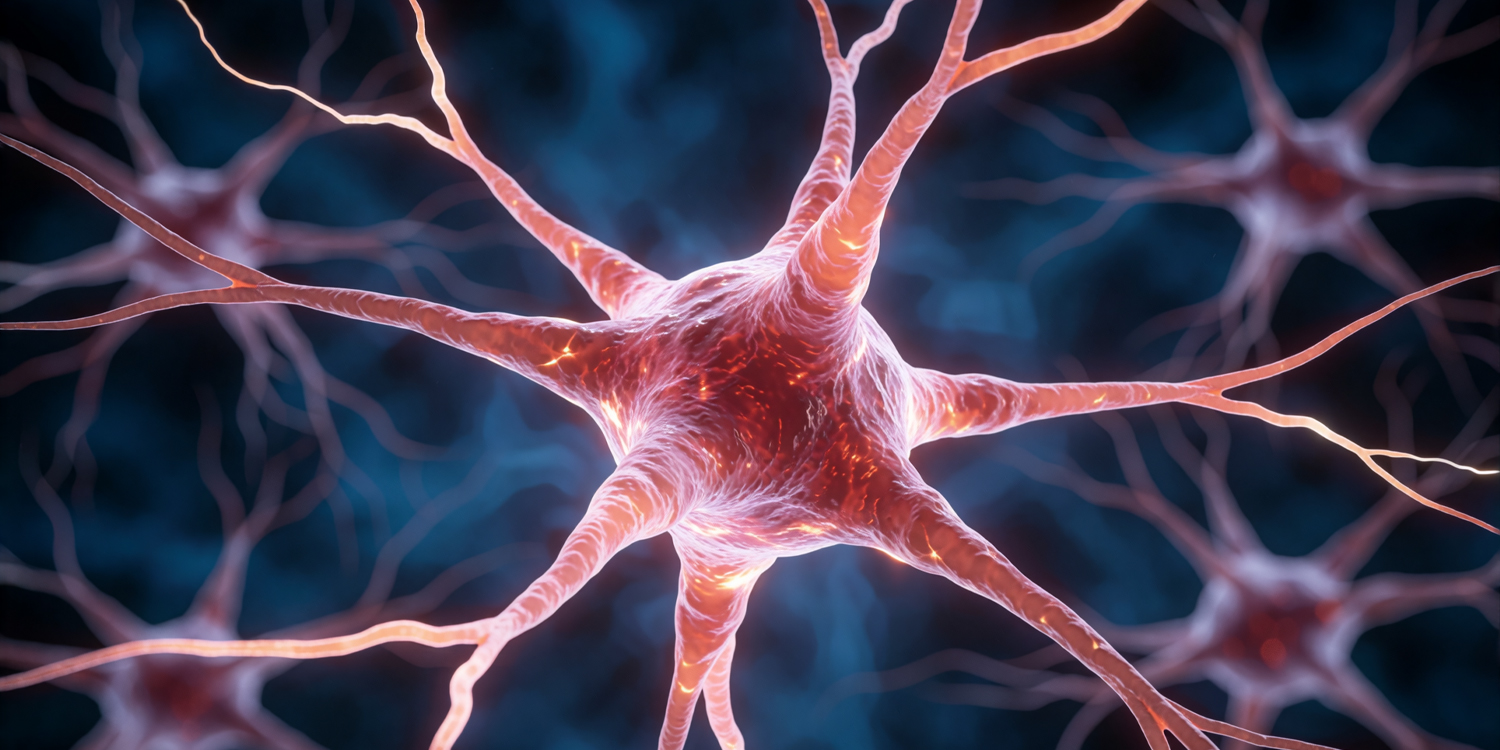
A new study reveals how antidepressant medications physically change the brain. Researchers found that treating chronic depression normalizes abnormal brain tissue microstructure, suggesting the initial abnormalities were actually the brain’s attempt to cope with severe symptoms.
An experimental study of individuals suffering from persistent depressive disorder found that treatment with the antidepressants duloxetine and desvenlafaxine normalized tissue microstructure in parts of the brain. Symptom severity mediated the treatment effects on tissue microstructure, suggesting that abnormal tissue at the start of the study might be a compensatory response of the brain to depression symptoms. The paper was published in NeuroImage: Clinical .
Persistent depressive disorder, formerly called dysthymia, is a chronic condition in which low mood and related symptoms persist for at least two years in adults. Symptoms may include low energy, poor concentration, hopelessness, low self-esteem, sleep disturbance, appetite changes, and reduced enjoyment or motivation. These symptoms may be less intense than those of a major depressive episode, but their long duration can substantially impair work, relationships, and quality of life.
Some people with persistent depressive disorder also experience superimposed episodes of major depression, sometimes informally called “double depression.” The mainstream treatments involve psychotherapy, medication, or a combination of both. Study
Source and reference
author Ravi Bansal, a researcher affiliated with the University of Southern California and Children’s Hospital Los Angeles, and colleagues note that previous studies have reported changes to the microstructure of different cortical and subcortical regions of the brain in individuals suffering from persistent depressive disorder. Studies have also noted that people who experience a reduction of depressive symptoms or remission in response to antidepressant medication also tend to see the structural changes associated with depression returning to normal. These, for example, include increased volume of the amygdala and hippocampus and thickening of the limbic cortex region of the brain in people who experience a remission or have milder clinical symptoms after treatment. In contrast, the volume of these areas declines and the cortex thins in individuals who do not remit or have more...
Read original source- Published
- Jul 11, 2026
- Updated
- Jul 11, 2026
- Source
- Psypost - Psychology News
- Category
- Health
- Read time
- 4 min
Key facts
Why this matters locally
This health story matters locally because it may affect readers, businesses, commuters, families, or public services in British Columbia.
Local impact
BC Post links this item to British Columbia coverage so readers can follow related city updates, weather, traffic, events, and category news in one place.
Timeline
Source and credit
BC Post may summarize, organize, and add local context for reader clarity. Original reporting remains with the listed publisher.
Related Articles
View all Health
Provation Expands Global Footprint with Launch of Provation® Apex in Australia and New Zealand
Health
Provation Expands Global Footprint with Launch of Provation® Apex in Australia and New Zealand
 Health
The Surprising Cellular Benefit of the Pigment Behind Red Hair
Health
The Surprising Cellular Benefit of the Pigment Behind Red Hair
 Health
Coffee May Protect the Liver in More Ways Than Scientists Realized
Health
Coffee May Protect the Liver in More Ways Than Scientists Realized
 Health
Study ties some birth control to brain tumours, but risks ‘rare’: experts
Health
Study ties some birth control to brain tumours, but risks ‘rare’: experts
 Health
Neuroscientists find a teamwork paradox: highly synchronized brains perform worse at complex tasks
Health
Neuroscientists find a teamwork paradox: highly synchronized brains perform worse at complex tasks
More Local Coverage
Latest news Politics
Powerful pro-life ad featuring boxing legend to air during final World Cup matches
Politics
Powerful pro-life ad featuring boxing legend to air during final World Cup matches
 Business
AI Market Trends: Q2 2026 Review and Forecast
Business
AI Market Trends: Q2 2026 Review and Forecast